Introduction
Although a small amount of sealer extrusion beyond the apical foramen is often considered a minor procedural mishap, extrusion into the inferior alveolar nerve (IAN) canal may lead to devastating complications, including persistent paresthesia, dysesthesia, or even permanent nerve injury. While such complications are relatively uncommon, they are largely preventable when appropriate precautions are taken during diagnosis, canal preparation, and obturation.
This article summarizes practical clinical recommendations that can significantly reduce the risk of sealer extrusion into the IAN canal.
1. Carefully Evaluate Preoperative Radiographs
The first step begins before initiating treatment.
Whenever the root apex of a mandibular premolar or molar appears to be in contact with the inferior alveolar canal, clinicians should assume that the relationship is genuine until proven otherwise.
It is often tempting to attribute the appearance to image superimposition or projection errors. However, in these situations, the safest approach is to assume close anatomical proximity and modify the treatment accordingly. Whenever there is doubt, always choose the safer clinical approach.
2. Establish an Accurate Working Length
Accurate working length determination is essential.
Electronic apex locators should be routinely used, even when high-quality radiographs are available. The working length should also be re-evaluated whenever necessary during treatment, particularly after coronal enlargement or when patency has been re-established.
Equally important is preservation of the apical constriction during cleaning and shaping. Excessive apical enlargement or destruction of the apical stop substantially increases the likelihood of sealer extrusion.
3. Sealer Selection Matters
Injectable bioceramic sealers are highly flowable and possess a strongly alkaline pH, frequently approaching 12 during the initial setting reaction. Although these materials demonstrate excellent biological properties inside the root canal system, extrusion into the inferior alveolar canal may expose neural tissues to chemical irritation and potentially contribute to nerve necrosis.
Therefore, if you choose to use a bioceramic sealer in these high-risk cases, never inject it directly into the root canal. Instead, follow the sealer placement technique presented later in Tip #6 for a safer and more controlled application.
For high-risk cases, a low-flow resin-based sealer may provide greater control during obturation.
Avoid using zinc oxide-eugenol (ZOE) sealers because the eugenol component can be highly irritating and neurotoxic to neural tissues if extruded into the IAN canal.
4. Use the Minimum Effective Amount of Sealer
A common misconception is that increasing the volume of sealer improves the quality of obturation.
In reality, excess sealer only increases the risk of extrusion without improving the seal.
In cases in close proximity to the IAN, only a minimal amount of sealer should be introduced into the canal.
5. Intentionally Position the Master Cone Slightly Short
Cleaning and shaping should always be completed to the full working length.
However, in teeth with a high risk of inferior alveolar canal involvement, the master cone may intentionally be positioned approximately 1-2 mm short of the working length while maintaining a distinct tug-back (Figure 1).
This strategy helps minimize the risk of forcing sealer beyond the apex.
6. Coat the Master Cone Properly
Rather than heavily coating the gutta-percha cone, only a very thin film of sealer should be applied.
Importantly, the apical 1–2 mm of the master cone should remain completely free of sealer.
This simple modification substantially decreases the amount of sealer delivered to the apical foramen.
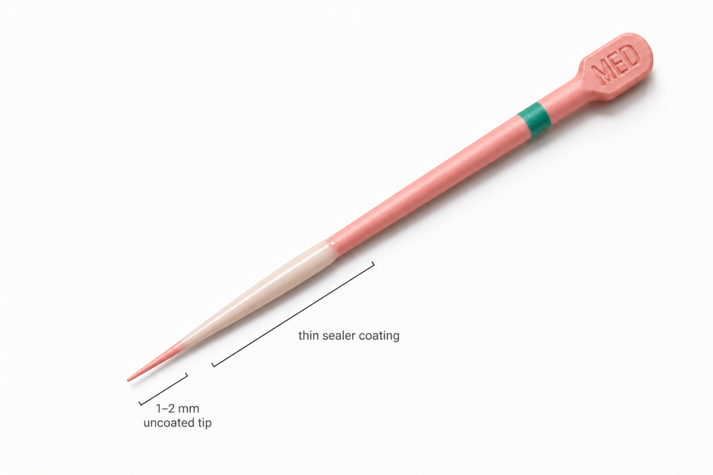
(Figure 1) Courtesy of Dr. M. Kalantar Motamedi
7. Insert the Cone Slowly
The master cone should be inserted slowly and smoothly.
Once the cone reaches its final position, repeated pumping or up-and-down movements should be avoided because they may hydraulically force sealer through the apical foramen.
Gentle insertion minimizes hydraulic pressure and reduces extrusion.
8. Technique-Specific Precautions
When performing lateral condensation, accessory cones should be marked with a reference point to avoid inadvertent overextension.
When using warm obturation techniques, the penetration depth of the heat carrier should be carefully controlled. Excessive penetration may generate sufficient hydraulic pressure to push both gutta-percha and sealer beyond the apex.
Carrier-based obturation technique should be avoided.
9. An Important Clinical Pearl
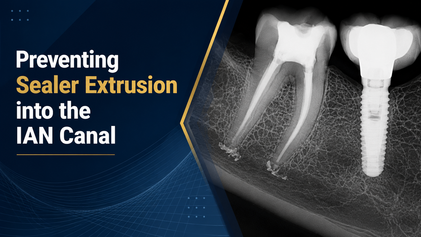
Clinical experience demonstrates that sealer may still reach the mandibular canal despite an apparent radiographic separation between the root apex and the canal (Figure 2).
This phenomenon is particularly relevant in teeth presenting with periapical lesions.
Unlike cortical bone, the bone surrounding the inferior alveolar canal is largely cancellous and contains interconnected trabecular spaces. Extruded sealer may therefore migrate through these marrow spaces before eventually entering the inferior alveolar canal.
Consequently, clinicians should exercise the same degree of caution in teeth with significant periapical bone loss, even when the radiograph appears to show a safe distance from the mandibular canal.
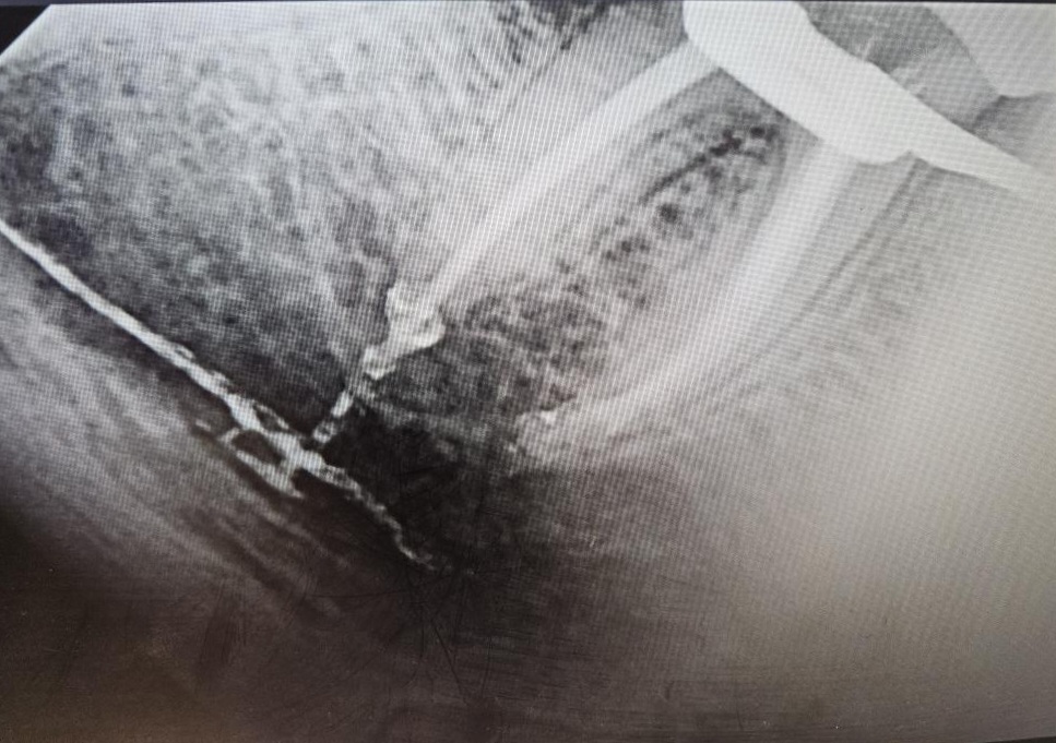
(Figure 2). This is a case from a dental school (courtesy of Dr. M. Kalantar Motamedi).
Conclusion
Sealer extrusion into the inferior alveolar canal is an uncommon but potentially life-altering complication. Fortunately, most cases can be prevented through meticulous diagnosis, accurate working length determination, preservation of the apical constriction, conservative sealer application, controlled master cone placement, and gentle obturation techniques.
Ultimately, prevention remains the best treatment for inferior alveolar nerve injuries associated with endodontic therapy.